
 TR
TR EN
EN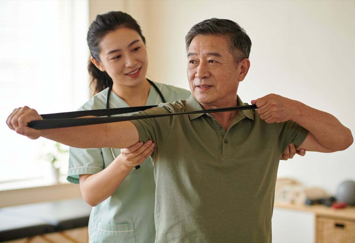
Overview
The menisci are two C-shaped cartilage structures in our knee joint, located between the thigh bone (femur) and the shin bone (tibia). We have two menisci, an inner (medial) and an outer (lateral) one. These structures are important tissues that distribute the load placed on the knee, stabilize the joint and act as shock absorbers.
A meniscus tear occurs when these cartilage cushions are damaged as a result of a sudden strain or wear that develops over time. The location, shape and size of the tear directly affect the treatment approach.
Causes
- Sudden twisting movements — a sudden twist of the knee while the foot is fixed is a common cause, especially in athletes.
- Degenerative wear — with age the meniscus tissue thins and loses its elasticity; it can tear even with minor strain.
- Sudden squatting and rising — straining the knee in deep flexion can lead to a tear.
- Excess weight — increases the load on the knee and accelerates wear of the meniscus tissue.
- Muscle weakness — weakness of the thigh (quadriceps) and hamstring muscles leaves the knee unprotected and prone to strain.
Symptoms
- Pain localized to the inner or outer side of the knee
- A sensation of swelling and fullness in the knee
- A feeling of catching, locking or giving way during movement
- Difficulty and pain when squatting and going up and down stairs
- Restriction in fully straightening or fully bending the knee
- A clicking sound heard with certain movements
Diagnosis
Physical examination is essential in diagnosing a meniscus tear. Our doctor listens to the patient's history, identifies the location of the pain and applies meniscus-specific clinical tests (such as McMurray and Apley). These tests provide important clues about the presence and location of the tear.
MRI is a valuable method for clarifying the type and size of the tear. However, it must be remembered that imaging alone is not sufficient; in many people meniscus wear may be seen on MRI without any complaints at all. For this reason, imaging findings must always be evaluated together with the physical examination.
Treatment
A significant proportion of meniscus tears, especially degenerative tears and those in the outer region, can be managed with conservative methods, without the need for surgery. We regard surgery as a last resort. The conservative treatment options are as follows:
- Rest and load adjustment — reducing the load on the knee in the acute phase.
- Physical therapy — reducing pain and swelling, then strengthening the muscles around the knee.
- Manual therapy and mobilization — achieving joint mobility and muscle relaxation.
- Personalized exercise program — protecting the joint by strengthening the muscles surrounding the knee.
What We Do
When a patient comes to us with knee pain, our doctor first performs a detailed physical examination; by evaluating the clinical tests and imaging findings together, they make the diagnosis and draw up a personalized treatment protocol. We raise surgery only in cases where it is essential.
In the phase where pain and swelling are pronounced, our doctor supports the process with dry needling and injection applications when deemed appropriate. Our specialist physiotherapists then relax the tense muscles around the knee with mobilization, cupping therapy and clinical massage.
Once relief is achieved, we move on to our main goal: creating the natural muscular support that protects the knee. With fully personalized, three-dimensional exercises, we strengthen the thigh and hip muscles. Because the human body is three-dimensional; we apply personalized rather than standard exercise.
We finish our sessions with kinesiology (athletic) taping so that the treatment is carried throughout the day. Throughout the process we follow our client closely and see them off with full satisfaction. Our principle is clear: we treat the person, not just the condition.